


Soundness is the currency of equine athletic longevity. Whether navigating Olympic grand prix dressage tests, sliding across reining pens, or quietly exploring mountain trails, an equine athlete relies on synovial articulations operating under immense mechanical strain. Establishing a proactive strategy for equine joint health care preserves articular cartilage, mitigates chronic inflammation, and extends your horse’s active career by years.
Joint degeneration-broadly categorized as osteoarthritis or degenerative joint disease (DJD)-is an insidious process. By the time visual lameness or joint effusion appears, irreversible micro-structural damage to the hyaline matrix has already occurred. In this comprehensive guide, we dissect the anatomy of the equine synovial joint, clinical lameness diagnostics, oral nutraceutical trials, injectable therapies, and progressive 16-week rehabilitation schedules.
Table of Contents
1. Anatomy of the Equine Synovial Joint
To execute effective equine joint health care, equestrians must understand how a movable synovial joint functions under athletic loading:
- Articular (Hyaline) Cartilage: A glassy, low-friction tissue covering the ends of articulating bones. Composed of specialized chondrocytes embedded in an extracellular matrix of type-II collagen and water-binding proteoglycans. Cartilage has zero blood supply or nerves-it relies entirely on joint fluid compression for nutrient diffusion.
- Synovial Fluid & Membrane: The inner lining (synovial membrane) secretes viscous, golden synovial fluid rich in high-molecular-weight hyaluronic acid (HA) and lubricin. This fluid lubricates articular surfaces and delivers metabolic nutrients.
- Subchondral Bone: The dense osseous layer directly supporting the cartilage cap. Intense concussive overload causes microfractures and sclerosis in subchondral bone, transmitting damaging forces upward into the cartilage.
2. High-Risk Joints: Hock, Stifle, and Fetlock Vulnerabilities
Different disciplines predispose specific anatomical joints to wear and tear:
- The Hock (Distal Tarsus): Bone spavin (osteoarthritis of the distal intertarsal and tarsometatarsal joints) is the most frequent hindlimb lameness in jumping and dressage horses. Because these lower joints have minimal range of motion, therapeutic fusion often resolves discomfort permanently.
- The Stifle (Femoropatellar & Femorotibial): The equine equivalent of the human knee. Meniscal tears and cruciate ligament strain occur frequently in performance cutting, jumping, and eventing horses.
- The Fetlock (Metacarpophalangeal Joint): Sustains extreme hyperextension during galloping and landing. High concussive loading predisposes to chronic synovitis, osteochondral fragments (chips), and sesamoiditis.
3. Early Warning Signs of Joint Inflammation and Arthritis
Early identification prevents acute synovitis from escalating into permanent cartilage erosions:
- Warm-Up Stiffness: A horse that feels wooden, short-strided, or resistant during the initial 10 to 15 minutes of trotting, but gradually works out of the stiffness as body temperature rises.
- Joint Effusion (Windpuffs & Bog Spavin): Visible swelling or fluid accumulation within the joint capsule (e.g., fetlock windpuffs or tibiotarsal hock swelling) that feels spongy or tight.
- Reluctance to Bend or Collect: Difficulty striking off on a specific canter lead, hollow transitions, tail swishing during downward transitions, or resistance when backing up.
- Uneven Shoe Wear: Inspect the underside of horseshoes or bare hooves. Uneven medial or lateral wear patterns indicate subtle gait compensation to offload an uncomfortable joint.
4. The Veterinary Lameness Exam: Flexion Tests and Blocks
Accurately diagnosing joint pain requires methodical veterinary examination:
- Passive Flexion Tests: The veterinarian manually holds a joint in extreme flexion for 60 seconds, then instructs the handler to trot the horse off immediately on a hard, level surface. Exaggerated head nodding or hip hiking indicates localized joint pain.
- Diagnostic Regional Nerve & Joint Blocks: Injecting local anesthetic (mepivacaine) systematically desensitizes specific neural pathways. If a 50% lame horse suddenly trots 100% sound after a coffin joint block, the lesion is confirmed inside that capsule.
- Diagnostic Imaging: Digital radiographs detect bone spurs (osteophytes) and joint space narrowing. Diagnostic ultrasound visualizes collateral ligaments, while Standing MRI reveals cartilage thinning months before bone changes appear.
5. The Science of Joint Supplements: What Really Works?
The oral joint supplement market is crowded, but veterinary clinical trials validate key bioactive ingredients:
- Glucosamine & Chondroitin Sulfate: Fundamental amino sugars that serve as building blocks for glycosaminoglycans (GAGs). They inhibit inflammatory cytokines like interleukin-1 (IL-1) and protect existing cartilage matrix from enzymatic degradation.
- Hyaluronic Acid (Oral & Injectable): High-molecular-weight oral HA helps maintain healthy synovial viscosity. Intravenous forms (such as Legend / hyaluronate sodium) and intra-articular injections rapidly restore lubricant properties directly within the joint capsule.
- MSM (Methylsulfonylmethane): A bioavailable organic sulfur donor with potent anti-inflammatory and antioxidant properties that reduces oxidative muscle and connective tissue damage post-exercise.
- Omega-3 Fatty Acids (EPA & DHA): Marine algae or fish-derived Omega-3s effectively alter cell membrane phospholipid composition, reducing systemic prostaglandin E2 synthesis.
- Polysulfated Glycosaminoglycans (Adequan): The FDA-approved gold standard intramuscular therapy that actively penetrates synovial fluid to stimulate chondrocyte synthesis.
6. Joint Injections: Steroids vs. HA vs. Polyacrylamide Hydrogels
When systemic therapies are insufficient, targeted intra-articular injections deliver direct relief:
- Corticosteroids (Triamcinolone Acetonide / Betamethasone): Potent anti-inflammatories that rapidly eliminate synovitis. Low-dose triamcinolone actually exhibits chondroprotective properties, preserving proteoglycan synthesis.
- Hyaluronic Acid (High Molecular Weight): Frequently combined with steroids to restore lubrication and coat articulating chondrocytes.
- Polyacrylamide Hydrogels (Arthramid Vet / Noltrex): A breakthrough non-pharmaceutical synthetic gel. Unlike steroids that wash out, hydrogel integrates permanently into the synovial membrane, cushioning the joint and providing 12 to 24+ months of mechanical pain relief.
7. Exercise Biomechanics: Footing, Warm-Up, and Cryotherapy
How you manage conditioning determines cartilage longevity:
- A Strict 15-Minute Dynamic Warm-Up: Synovial fluid is thixotropic-it becomes less viscous and lubricates far more efficiently as temperature rises. Walking actively for 10 to 15 minutes warms the joint fluid, allowing cartilage to absorb shock before intense collection or jumping begins.
- Surface Footing Quality: Deep, uncompacted sand places extreme torque on soft-tissue suspensory ligaments and distal joints. Hard, unyielding clay transmits severe concussive shock waves straight up the pastern and coffin joints. Maintain level, elastic, dust-free arena footing.
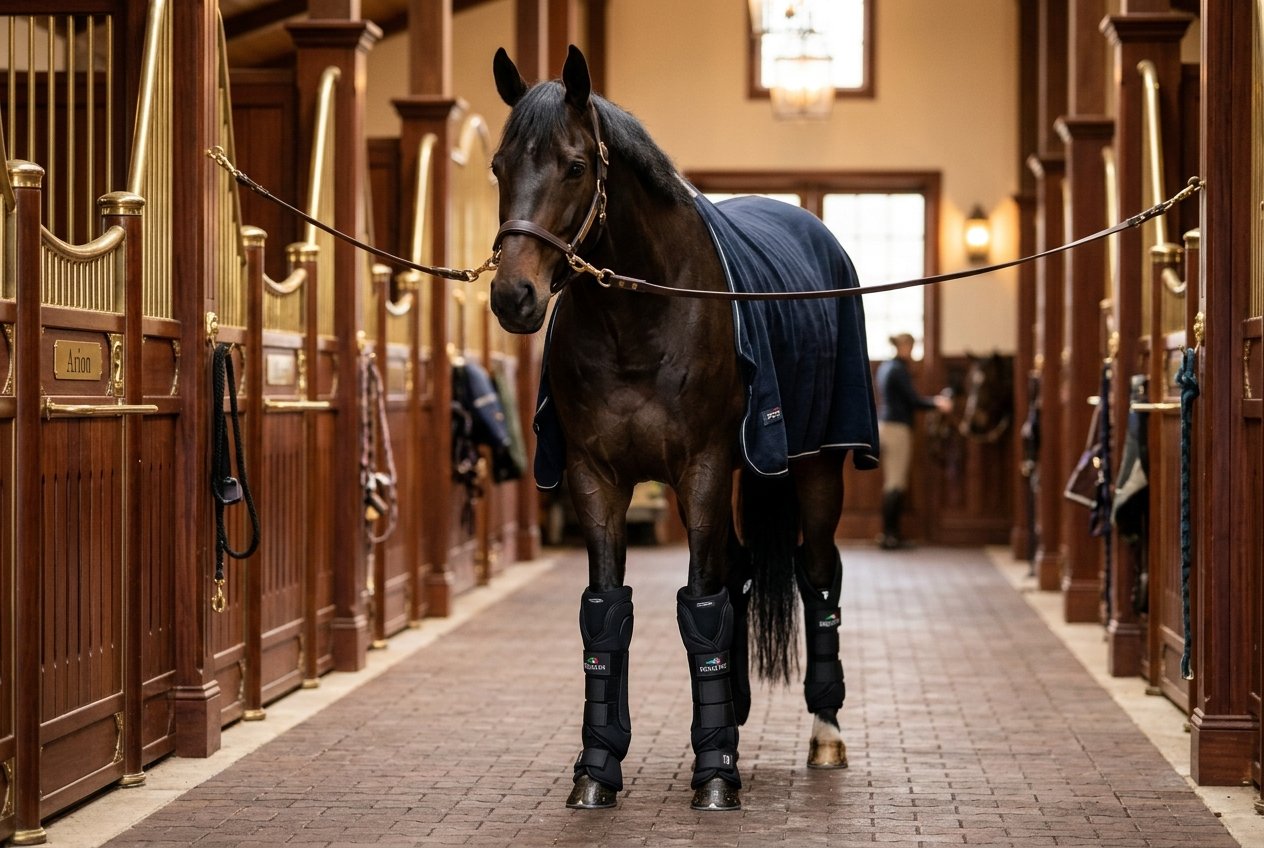
- Cold Therapy (Cryotherapy): Standing in ice boots or cold-hosing legs for 15 to 20 minutes post-workout constricts dilated capillaries, suppressing cellular inflammatory cascades before they damage synovial membranes.
8. Advanced Regenerative Medicine: PRP, IRAP, and Stem Cells
When structural cartilage degradation progresses, advanced equine sports medicine offers revolutionary regenerative protocols:
- IRAP (Interleukin-1 Receptor Antagonist Protein): Blood is drawn from the horse, incubated with medical-grade glass beads to amplify anti-inflammatory cytokines, and injected back into the joint to neutralize destructive enzymes.
- PRP (Platelet-Rich Plasma): Concentrated autologous platelets release massive levels of growth factors (TGF-beta, PDGF) to promote soft tissue and cartilage remodeling.
- Stem Cell Therapy: Bone marrow-derived or adipose-derived mesenchymal stem cells (MSCs) deliver cellular regeneration to severely damaged joint environments.
9. Structured 16-Week Rehabilitation Return-to-Work Protocol
Returning a horse to work after a joint flare-up requires progressive loading:
- Weeks 1 to 4: Hand walking only, starting at 10 minutes daily on firm, flat footing and progressing to 30 minutes twice daily. Cold therapy after every session.
- Weeks 5 to 8: Introduce under-saddle walk work (30 to 40 minutes). Begin gentle straight-line trot sets (2 minutes trot, 5 minutes walk). Avoid tight circles and small lunging rings.
- Weeks 9 to 12: Increase trotting to 15 minutes total. Introduce wide, flowing 20-meter circles. Begin controlled straight canter sets on good arena footing.
- Weeks 13 to 16: Progressive sport-specific collection, lateral work, or low cavaletti, with veterinary soundness check before full competition resumption.
Frequently Asked Questions
At what age should I start giving my horse joint supplements?
Starting early is far more effective than trying to reverse existing damage. Horses entering consistent training at age 3 to 5 benefit enormously from preventative oral GAGs and high-molecular-weight hyaluronic acid.
Do joint injections ruin the joint over time?
When administered judiciously under veterinary guidance, modern low-dose corticosteroid formulations reduce damaging joint inflammation without compromising cartilage metabolism. Over-injecting without rest, however, can mask pain and lead to accelerated structural wear.
Can turn-out in pasture help arthritic horses?
Yes! Continuous 24/7 turnout with gentle walking motion keeps synovial fluid constantly circulating, preventing the severe stiffness that occurs when arthritic horses stand confined in a stall for 20+ hours daily.
Conclusion
Comprehensive equine joint health care requires an intelligent synergy between daily stable management, surface conditioning, evidence-based nutrition, and early veterinary intervention. By protecting cartilage before irreversible degradation takes hold, you empower your equine athlete to move with fluidity, joy, and effortless power throughout every phase of life.
