

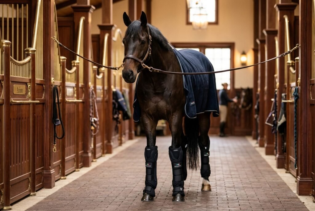
In every equestrian discipline—from high-performance Olympic grand prix jumping to leisurely weekend trail riding—soundness is the irreplaceable bedrock of equine performance and welfare. A subtle hitch in a horse’s stride, an almost imperceptible head nod at the trot, or a reluctance to step under the body in canter are rarely manifestations of behavioral stubbornness; rather, they are early biological distress beacons signaling physical discomfort. This comprehensive horse lameness detection guide equips owners, riders, and trainers with the clinical diagnostic knowledge to identify asymmetrical gait irregularities, evaluate limb biomechanics, understand veterinary diagnostic nerve blocks, and navigate modern regenerative rehabilitation therapies.
🐎 The AAEP Official Lameness Scale Reference Card
Table of Contents
1. The Biomechanics of Asymmetry: Reading the Equine Gait
A completely sound horse in motion displays symmetrical rhythm, equal ground-contact duration across bilateral pairs of limbs, and smooth vertical oscillations of the head and pelvis. The trot is universally selected by veterinarians as the primary diagnostic gait because it is a symmetrical, two-beat diagonal gait (left foreleg and right hindleg move forward synchronously, alternating with right foreleg and left hindleg). At the trot, there is no suspension moment between diagonal pairs that could mask weight transfer.
When a limb experiences pain upon impact or propulsion, the horse instinctively modifies its kinematics to minimize ground reaction forces on that injured leg. This mechanical compensation manifests as subtle vertical displacement shifts that the trained human eye can decipher.
2. Forelimb vs. Hindlimb Lameness: The Axioms of Motion
The golden rules of visual lameness identification differ significantly between front and hind limbs:
The Forelimb Rule (“Down on Sound”):
When a horse has a sore front leg, it uses the heavy mass of its head and neck (which acts as a pendulum counterweight) to relieve concussion. As the lame foreleg impacts the ground, the horse raises its head and neck upward. Conversely, when the opposite, pain-free foreleg strikes the ground, the head and neck nod downward. Hence, the universal veterinary mnemonic: “Down on sound.” If the head goes up when the right front hits, the horse is lame on the right front.
The Hindlimb Rule (“Hip Hike & Drop”):
Identifying hindlimb lameness requires observing the horse trotting away from you in a straight line, focusing on the tubera sacrale (the highest points of the croup). As the lame hind foot strikes the ground, the gluteal muscles contract defensively, causing the hip on the lame side to hike upward (or demonstrate a markedly reduced downward excursion). Furthermore, the lame hind foot frequently tracks short (landing behind the print of the forefoot) and drifts toward the midline of the body.
3. The 5-Step Clinical Barn Exam: Digital Pulse, Palpation & Hard Surface Trot
Before calling the veterinarian, execute this methodical physical assessment:
- Palpating the Digital Pulse: Place your index and middle fingers over the neurovascular bundle running across the abaxial surface of the sesamoid bones at the fetlock. In a healthy, non-inflamed hoof, the digital pulse is faint or barely perceptible. A bounding, throbbing digital pulse combined with heat in the hoof capsule is a hallmark indicator of acute subsolar abscess or life-threatening laminitis.
- Systematic Leg Palpation: Run your hands down each limb while unweighted and weight-bearing. Feel for localized heat, edema (swelling), windpuffs in the digital flexor tendon sheath, thickening of the superficial digital flexor tendon (the “bowed tendon” profile), or pain upon palpation of the suspensory ligament branches.
- Hoof Tester Application: Systematically apply hoof testers across the sole, bars, and across the frog to detect focal pain indicative of bruising, corns, or navicular bursa irritation.
- Hard Surface Evaluation: Trot the horse in hand on a firm, level asphalt or packed gravel surface. Hard surfaces accentuate concussive pain in the lower hoof, ringbone, and bone spavins.
- Lungeing on Circles: Lunge the horse on both left and right 15-meter circles in a soft arena. Traveling in a circle increases torque on collateral ligaments and joints. Forelimb lameness is almost always exacerbated when the lame leg is on the inside of the circle.
4. Veterinary Flexion Tests: Stressing the Joints for Diagnosis
A flexion test involves manually holding a specific joint in deep, sustained flexion for 60 to 90 seconds, followed immediately by having the handler trot the horse briskly away in a straight line. By temporarily compressing articular cartilage and stretching joint capsules and tendons, flexion tests temporarily amplify low-grade subclinical pain. A horse that trots off with marked asymmetry after distal limb flexion (fetlock, pastern, coffin joint) directs the veterinarian’s diagnostic attention to the lower limb.
5. Diagnostic Analgesia: How Nerve and Joint Blocks Localize Pain
Because multiple structural issues can appear on X-rays without causing actual pain, veterinarians use perineural anesthesia (nerve blocks) to definitively pinpoint the anatomical source of discomfort. Using small doses of local anesthetic (carbocaine / mepivacaine), the vet works systematically from the bottom of the foot upward:
| Diagnostic Nerve Block | Anatomical Injection Site | Desensitized Zone | Target Pathology Identified |
|---|---|---|---|
| Palmar Digital (PD) Block | Over the palmar digital nerves at the collateral cartilages of the foot. | Palmar 1/3 of foot: navicular bone, bursa, DDFT insertion, sole corium. | Navicular syndrome, heel pain, subsolar abscess, deep digital flexor tendinitis. |
| Abaxial Sesamoid Block | Over the abaxial aspect of the proximal sesamoid bones. | Entire foot, pastern joint, distal sesamoidean ligaments, coffin joint. | Ringbone, collateral desmitis, extensive solar trauma, laminitis. |
| Low Four-Point (Low Palmar) | Just above the fetlock joint between the flexor tendons and suspensory branches. | Fetlock joint, sesamoid bones, digital flexor tendon sheath. | Fetlock synovitis, sesamoiditis, suspensory branch desmitis. |
6. Advanced Imaging: Digital Radiography, Ultrasound & MRI
Once nerve blocks localize the pain to a specific region, imaging technologies visualize the exact tissue damage:
- Digital Radiography (DR): Evaluates bony architecture, joint space narrowing, subchondral bone cysts, osteophytes (arthritis spurs), and pedal osteitis.
- High-Resolution Musculoskeletal Ultrasound: Evaluates soft tissue structures, revealing fiber disruption, cross-sectional tear size, and core lesions in the superficial and deep digital flexor tendons and suspensory ligament.
- Standing Low-Field MRI: The gold standard for foot lameness. It visualizes micro-fractures, cartilage loss, and soft-tissue injuries deep inside the hoof capsule that are completely invisible on standard X-rays and ultrasound.
7. Cutting-Edge Regenerative Therapies: PRP, IRAP, Shockwave & Stem Cells
Equine sports medicine has advanced beyond simple steroid injections. Modern rehabilitation utilizes autologous biological therapies that stimulate tissue regeneration rather than merely masking inflammation:
Platelet-Rich Plasma (PRP): Concentrated blood platelets harvested from the horse’s jugular vein contain potent transforming growth factors (TGF-beta, PDGF). When injected under ultrasound guidance directly into tendon and ligament tears, PRP promotes organized, aligned collagen synthesis, dramatically reducing reinjury rates.
IRAP (Interleukin-1 Receptor Antagonist Protein): Blood is incubated with specialized medical beads that stimulate production of natural anti-inflammatory proteins. Injected into arthritic joints, IRAP blocks the destructive cytokine Interleukin-1, halting cartilage breakdown without the catabolic side effects of repeated corticosteroid use.
Extracorporeal Shockwave Therapy (ESWT): Acoustic high-energy sound waves penetrate deep tissues, stimulating microcirculation, recruiting local progenitor cells, and providing prolonged localized analgesia in chronic suspensory desmitis and sacroiliac disorders.
8. Frequently Asked Questions (FAQ)
Why does my horse look sound at the walk, but lame at the trot?
The walk is a four-beat gait without a moment of suspension, which allows the horse to distribute its body weight evenly across three grounded feet at almost all times. The trot forces each diagonal pair of legs to absorb the entire gravitational mass of the horse, immediately revealing unilateral weaknesses.
How long does a tendon tear take to heal in a horse?
Equine tendons heal very slowly due to their limited vascular blood supply. A significant superficial digital flexor tendon lesion requires a minimum of 9 to 12 months of structured, ultrasound-monitored hand walking and progressive rehabilitation before returning to full athletic work.
📖 Recommended Soundness & Equine Health Reading
Explore more veterinary guides and joint care masterclasses across Horse Silk:
- Horse Silk Soundness Guide: Equine Joint Health & Longevity Care: Supplements, Injections & Management
- Horse Silk Hoof Care: Essential Horse Hoof Care 101: Anatomy, Daily Maintenance & Thrush Prevention
- Official Veterinary Standard: American Association of Equine Practitioners (AAEP) Lameness Scale
- Equine Research Portal: British Equine Veterinary Association (BEVA) Soundness Protocols
